
How Technology Makes The Result Precise
The problem
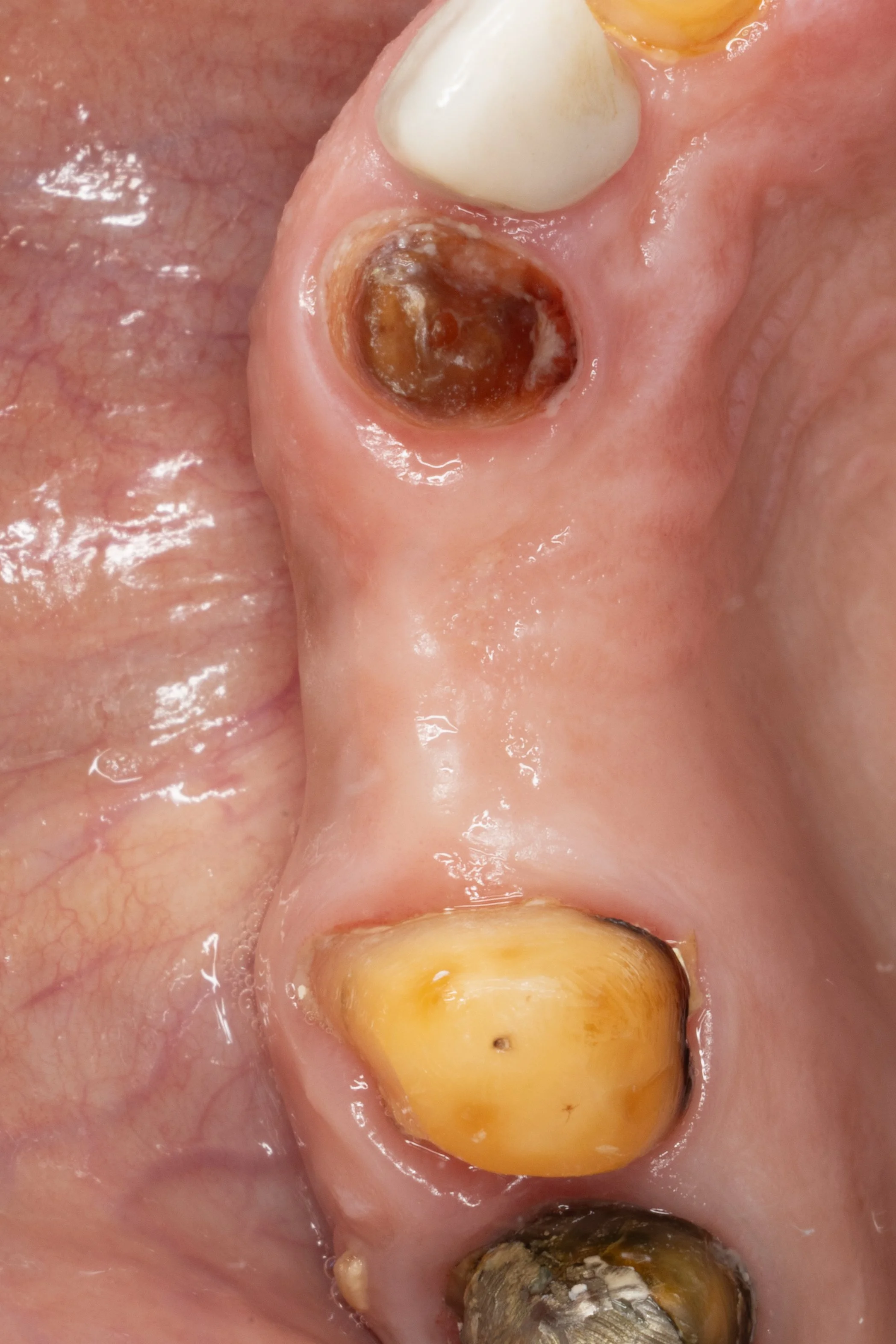
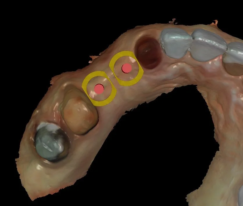
The patient came to our clinic because she was not comfortable with her old bridge on the upper left side. Two teeth were missing, and the bridge was fixed to the nearby teeth.
Over time, a space formed under the bridge. This caused:
Food getting stuck easily
Difficulty cleaning after meals
Inability to use dental floss
Gum discomfort
Why the old bridge was a problem
The patient came to our clinic because she was not comfortable with her old bridge on the upper left side. Two teeth were missing, and the bridge was fixed to the nearby teeth.
Over time, a space formed under the bridge. This caused:
Food getting stuck easily
Difficulty cleaning after meals
Inability to use dental floss
Gum discomfort
The best solution
We decided to:
Remove the old bridge
Place two dental implants to replace the missing teeth
Restore each tooth with individual crowns
This allows better cleaning, healthier gums, and a more natural result.
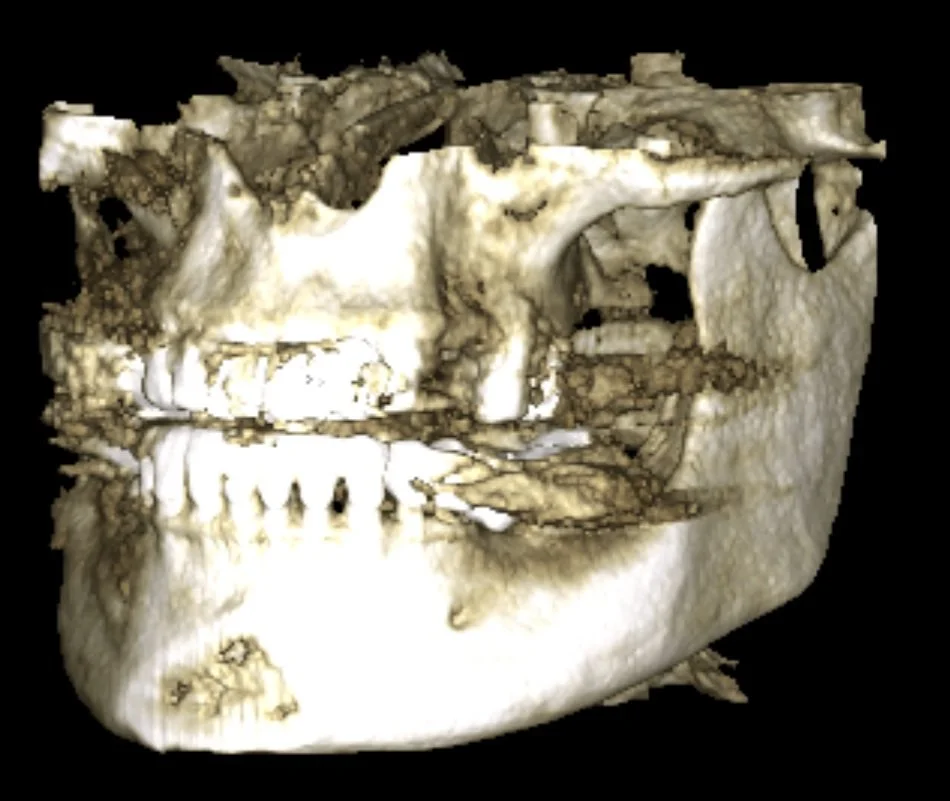
Step 1: Careful Examination & Planning
A 3D X-ray (CBCT) was taken to check the bone
We digitally designed the final shape of the new teeth
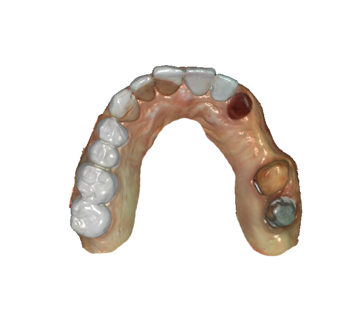
A digital scan of the teeth was done (no messy impressions)
The scan showed that the bone space was limited, so the implants had to be placed very accurately.
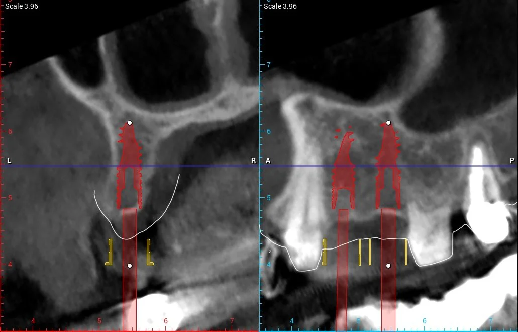
Step 2: Digital Surgery Preparation
Using specialized software, the CBCT and intraoral scans were merged into a single digital model. The missing teeth were digitally designed in their ideal position, and two high-quality implants were carefully planned according to the final crowns and available bone — without the need for bone grafting.
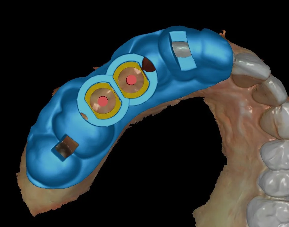
A customized digital surgical guide was then designed and 3D-printed using FDA-approved material to ensure exact implant placement during surgery.
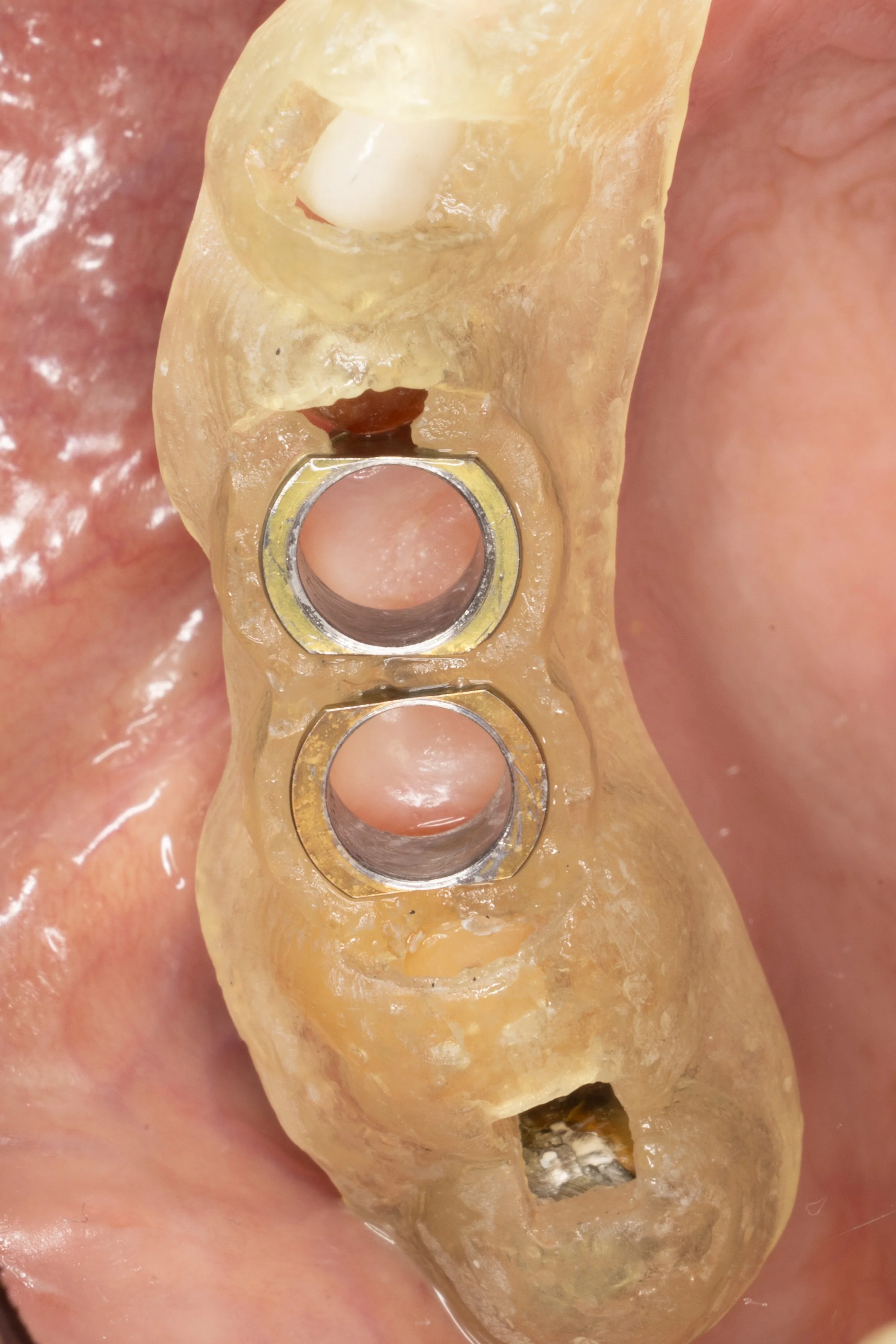
Step 3: Surgical Phase
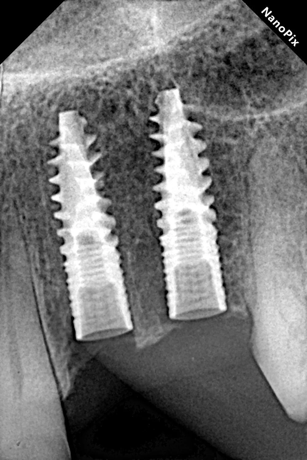
This phase is the easiest part of the whole procedure, as it is direct application of the previous one. The entire phase took only less than 30 minutes. Post-operative X-rays confirmed perfect implant positioning and parallelism.
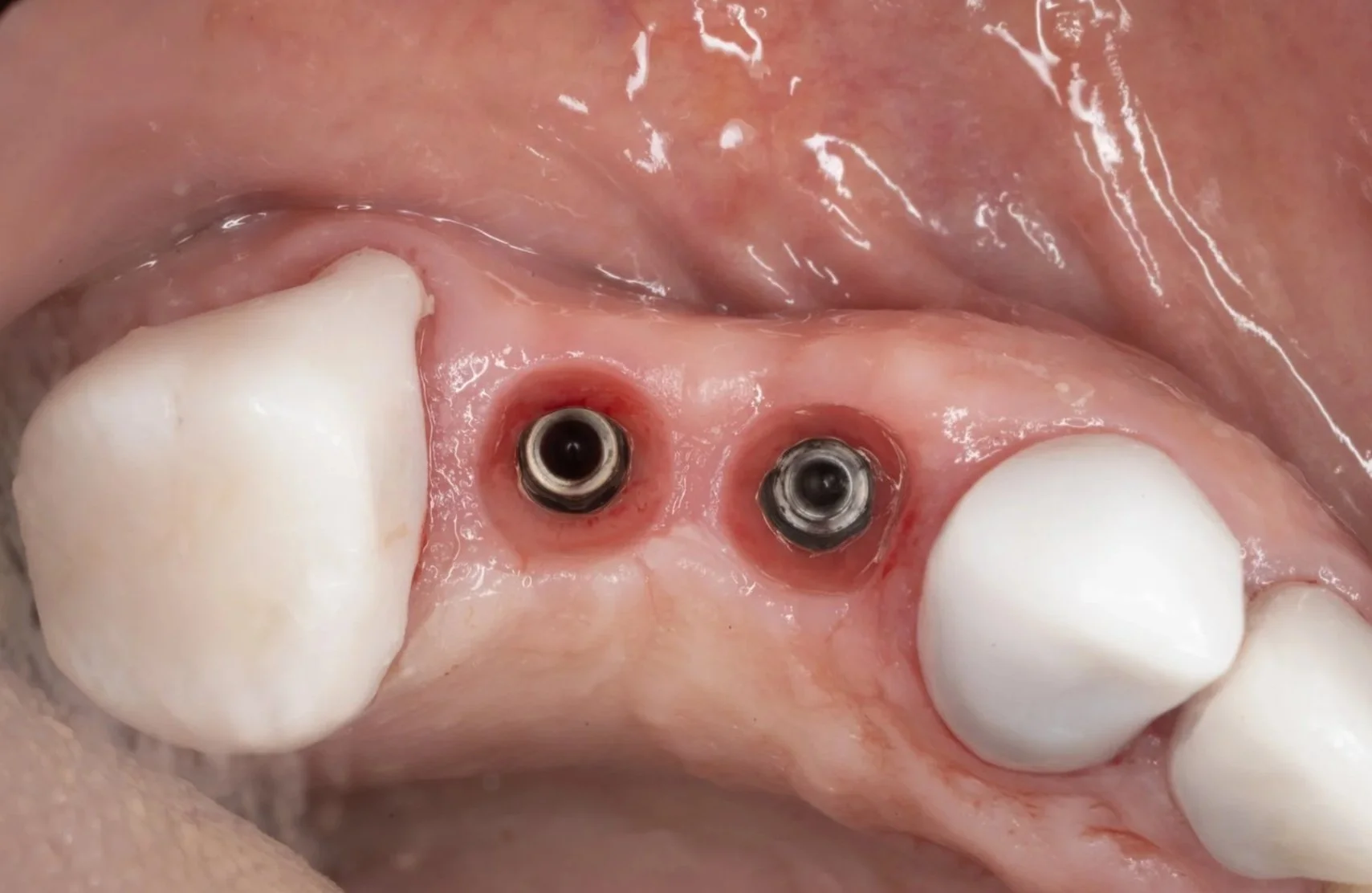
Step 3: Final Crowns
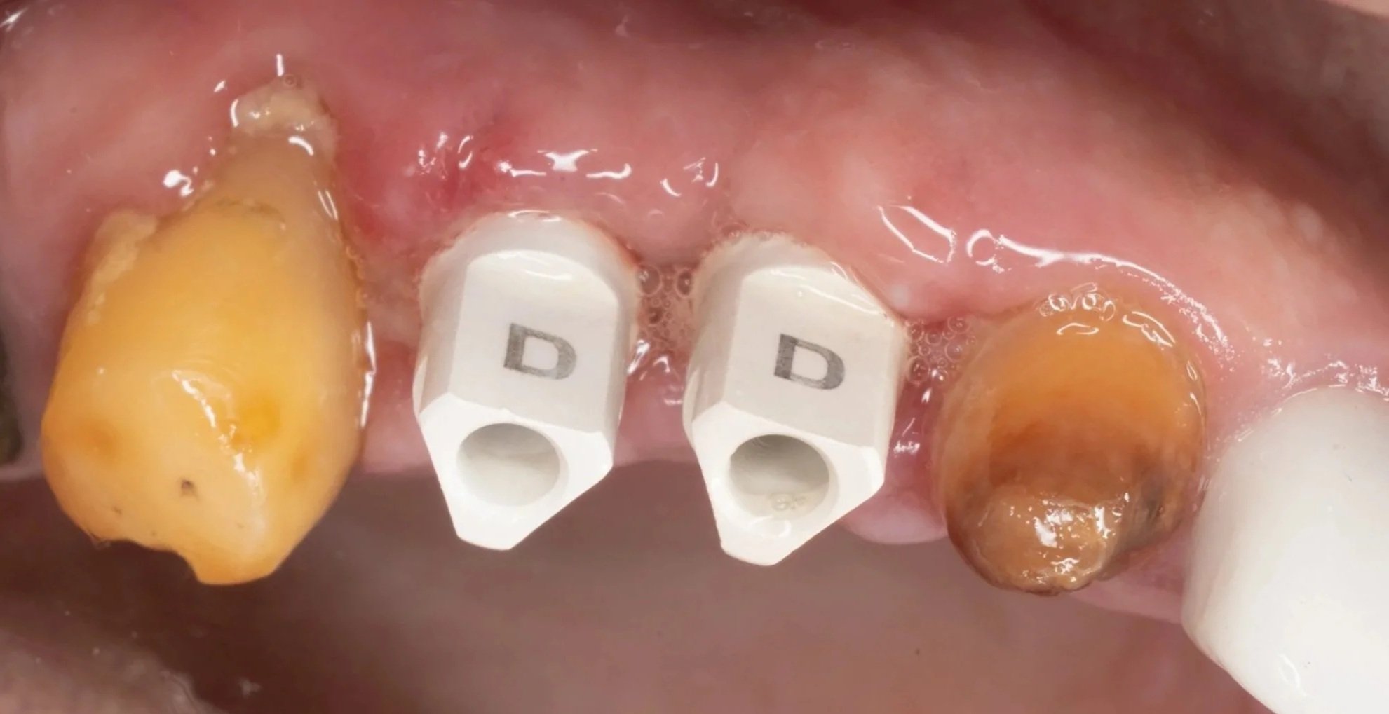
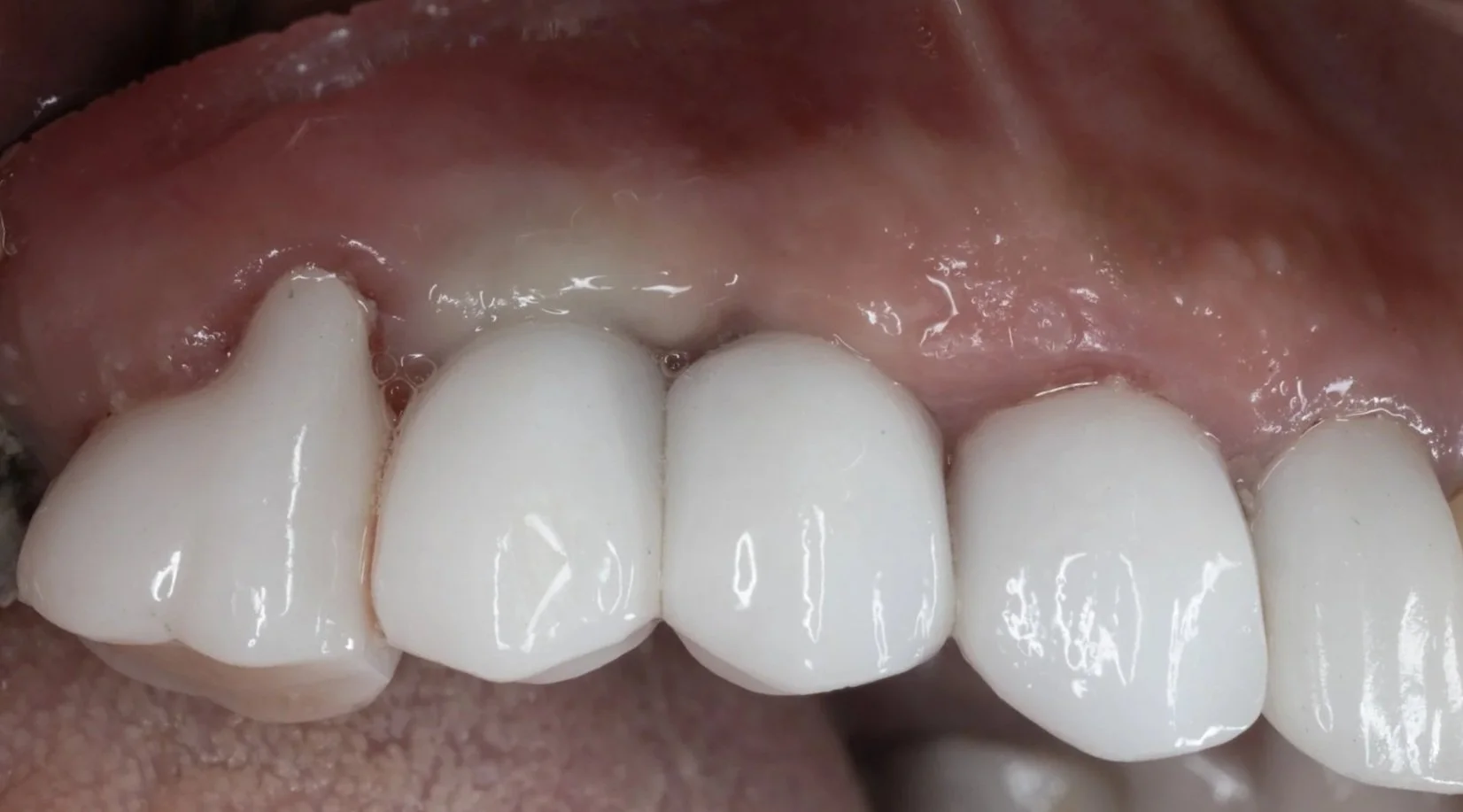
After three months of healing, digital impressions were taken using an intraoral scanner. The final crowns were delivered within one week, restoring both function and aesthetics. The patient was extremely satisfied with the result.
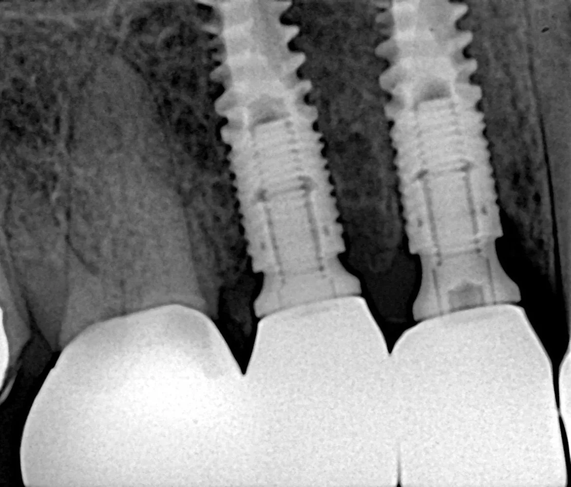
Precisely placed implants following international protocols
High-quality, natural-looking crowns with ideal gum relation
Healthy gums with easy cleaning and no food accumulation
This case highlights how digital technology allows maximum precision, predictable outcomes, and long-term oral health.